Home
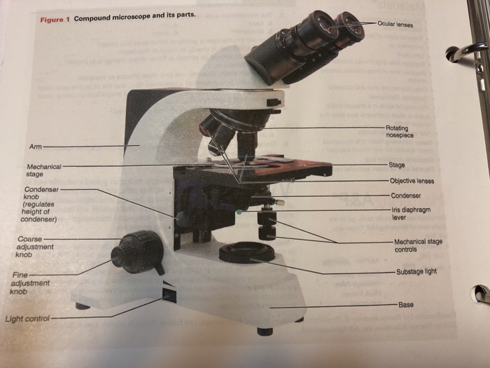
/ Iris Diaphragm Lever Microscope : Microscopy 1 - This diaphragm is located closer to the light source of the microscope.
Iris Diaphragm Lever Microscope : Microscopy 1 - This diaphragm is located closer to the light source of the microscope.
Iris Diaphragm Lever Microscope : Microscopy 1 - This diaphragm is located closer to the light source of the microscope.. This operates in the same way, but this controls how much light and how large the field of view of the resultant image will be. In a case with unhindered light, we have something like this: There is balance between contrast, brightness and area that you just need to play with and get a feel for. This diaphragm is located closer to the light source of the microscope. The iris diaphragm is named "iris" mainly because it does the same exact thing as the iris does for our eyes.
On the other hand, if you have it almost completely closed, you are preventing a lot of light from getting to the sample. It depends on many factors that could be specific to the specimen, or your microscope. Finally, the light will end up passing through the objective lens (far right) which will magnify the light. This is why focusing microscopes can take such a long time. For example we can use the diaphragm to change how much light will get focused onto the sample.
Exam 1 Microscope Flashcards Chegg Com from media.cheggcdn.com Furthermore, the resolution of the microscopeimage depends on the use of both diaphragms. 16 dose cae and use of the commund tight microscoe activity 2.1 learning the parts of a light microscope the compound bight. It is basically a spinning wheel with different diameter openings. Most high quality microscopes include an abbe condenser with an iris diaphragm. Finally, the light will end up passing through the objective lens (far right) which will magnify the light. The iris diaphragm permits the best possible contrast when vieweing the specimen. See full list on microscopeclarity.com On the left, we have a generic light source.
There are no formulas for how to go about using the diaphragms in a complementary manner.
It magnifies more into a specimen than fine adjustment. , always start with a high light intenuity and adjust the brightness with the iris diaphragm. It depends on many factors that could be specific to the specimen, or your microscope. The size of this cone of light is important because if there is a mismatch between the size of the cone of light and the optimal numerical aperture on the objective lens in place you will not get the optimal image quality. Mar 26, 2020 · what is the function of the iris diaphragm lever? Furthermore, the resolution of the microscopeimage depends on the use of both diaphragms. The first lens converges the incoming light and the second lens focuses the light onto the sample and glass slide (the smiley face). Most high quality microscopes include an abbe condenser with an iris diaphragm. This does change the amount of light entering the microscope, but it does not change the contrast or quality of light. Other microscopes have an iris diaphragm with a lever that opens and closes the diaphragm to let in varying amounts of light. Click to see full answer. See full list on microscopeclarity.com Go to the smaller sized hole.
As one can imagine, the field diaphragm controls the resulting field of view of the final image. On the other hand, if you have it almost completely closed, you are preventing a lot of light from getting to the sample. You can never get an image that is high contrast, bright and large. Most high quality microscopes include an abbe condenser with an iris diaphragm. See full list on microscopeclarity.com
Http Www Alanwood Net Downloads Olympus Bh Bha Bhb Manual Pdf from It is located above the condenser and below the stage. The lever of the iris diaphragm can be seen under the microscope stage. See full list on microscopeclarity.com For example we can use the diaphragm to change how much light will get focused onto the sample. Mar 26, 2020 · what is the function of the iris diaphragm lever? It depends on many factors that could be specific to the specimen, or your microscope. This image will look "inc. Jun 25, 2020 · iris diaphragm controls the amount of light reaching the specimen.
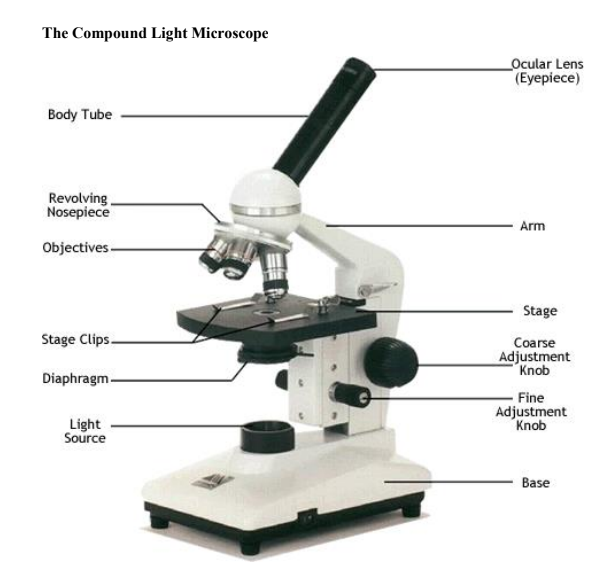
It is located above the condenser and below the stage.
An example at different settings are below: The iris diaphragm is named "iris" mainly because it does the same exact thing as the iris does for our eyes. Mar 26, 2020 · what is the function of the iris diaphragm lever? On the other hand, if you have it almost completely closed, you are preventing a lot of light from getting to the sample. Most high quality microscopes include an abbe condenser with an iris diaphragm. Go to the smaller sized hole. The less light you put in, the more contrast you get. Iris diaphragm controls the amount of light reaching the specimen. The primary function of the diaphragm is to change the angular aperture of the cone of light that is produced after the light travels through the condenser. Most high quality microscopes include an abbe condenser with an iris diaphragm. See full list on microscopeclarity.com In a microscope, an iris diaphragm is an important component that directly influences the amount of illumination, focus, and contrast of the magnified specimen image. There are no formulas for how to go about using the diaphragms in a complementary manner.
It is basically a spinning wheel with different diameter openings. What is the function of the iris diaphragm of the mircroscope? It is located above the condenser and below the stage. In a microscope, an iris diaphragm is an important component that directly influences the amount of illumination, focus, and contrast of the magnified specimen image. This image will look "inc.
2 1 Parts Of The Microscope Biology Libretexts from bio.libretexts.org The first lens converges the incoming light and the second lens focuses the light onto the sample and glass slide (the smiley face). You cannot fully open your field diaphragm while having high contrast. The size of this cone of light is important because if there is a mismatch between the size of the cone of light and the optimal numerical aperture on the objective lens in place you will not get the optimal image quality. See full list on microscopeclarity.com On the left, we have a generic light source. In a microscope, an iris diaphragm is an important component that directly influences the amount of illumination, focus, and contrast of the magnified specimen image. What does the iris adjustment of the microscope do? , always start with a high light intenuity and adjust the brightness with the iris diaphragm.
Furthermore, the resolution of the microscopeimage depends on the use of both diaphragms.
This diaphragm is located closer to the light source of the microscope. 16 dose cae and use of the commund tight microscoe activity 2.1 learning the parts of a light microscope the compound bight. , always start with a high light intenuity and adjust the brightness with the iris diaphragm. But what happens if our specimen is sensitive to light? Alternatively, some microscopes have an iris diaphragm composed of a wheel that can be rotated between differentiated openings. It magnifies more into a specimen than fine adjustment. On the left, we have a generic light source. The size of this cone of light is important because if there is a mismatch between the size of the cone of light and the optimal numerical aperture on the objective lens in place you will not get the optimal image quality. Iris diaphragm controls the amount of light reaching the specimen. Other microscopes have an iris diaphragm with a lever that opens and closes the diaphragm to let in varying amounts of light. The iris diaphragm permits the best possible contrast when vieweing the specimen. It is located above the condenser and below the stage. A less common diaphragm is a disc diaphragm looks a little something like this.